tree in bud opacities treatment
Tree-in-bud pattern seen on high-resolution CT HRCT indicates dilatation of bronchioles and their filling by mucus pus or fluid. 1 direct filling of the centrilobular arteries by tumor emboli and 2 fibrocellular intimal hyperplasia due to carcinomatous.
2
Video chat with a US.
. However to our knowledge the relative frequencies of the causes have not been evaluated. 11 TIB opacities represent a central imag- Background. The tree-in-bud pattern occurs commonly in patients with endobronchial spread of Mycobacterium tuberculosis and is highly suggestive of active tuberculosis 2 3High-resolution CT usually reveals small 24-mm centrilobular nodules and branching linear opacities of similar caliber originating from a single stalk Figs 2 3 4.
The latter etiology is often overlooked but is important to consider in patients with a cancer history to avoid delays in diagnosis and treatment. The tree-in-bud sign indicates bronchiolar luminal impaction with mucus pus or fluid causing normally invisible peripheral airways to become visible 80. Treatment aims to slow the progression of the condition.
Invasive airway aspergillosis should be suggested when the tree-in-bud pattern occurs in combination with a consolidation accompanied by a halo of ground-glass opacity in a patient with leukemia. Tree in bud opacities treatment. The purpose of this study was to.
In clinical practice however it can reflect a wide variety of pathogens including bacterial fungal and viral organisms. The most common CT findings are centrilobular nodules and branching linear and nodular opacities. Thus the bronchioles resemble a branching or budding tree and are usually somewhat nodular in appearance This morphologic pattern can be seen in a wide variety of diseases as illustrated by Gosset et al.
Cytomegalovirus pneumonia in a 51-year-old man with chronic myelogenous leukemia who underwent bone marrow transplantation. An 82yearold man was transferred to our emergency department with symptoms of chills and 405C fever. View larger version 145K Fig.
In the December 2009 issue of the AJR. However to our knowledge the relative frequencies of the causes have not been evaluated. 78 indicating the absence.
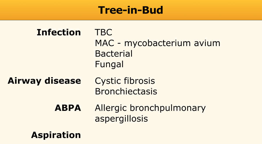
Treeinbud opacities detected after aspiration should be considered DAB rather than mycobacterial infection. The tree-in-bud pattern might be helpful to choose a suitable therapy in patients with an acid-fast bacilli smear-positive diagnosis of lung disease. 1 2 3 4 reported causes include infections aspiration and a variety of inflammatory conditions.
Get prescriptions or refills through a video chat if the doctor feels. It is not specific for a single disease entity but is a direct sign of various diseases of the peripheral airways and an indirect sign of bronchiolar diseases such as air trapping or sub. The purpose of this study was to determine the relative frequency of causes of TIB opacities and identify patterns of disease associated with TIB opacities.
Correlation with symptoms and attention on follow up to ensure resolution is suggested. Board-certified doctor 247 in less than one minute for common issues such as. Intravascular pulmonary tumor embolism often occurs in cancers of the breast liver kidney stomach prostate and ovaries and can lead to the tree-in-bud sign in HRCT 214.
Multiple causes for tree-in-bud TIB opacities have been reported. The tree-in-bud pattern can be an early sign of disease Fig 10 15. The tree-in-bud sign has been described in cases of acute aspiration 13.
However to our knowledge the relative frequencies of the causes have not been evaluated. Although initially described in 1993 as a thin-section chest CT finding in active tuberculosis TIB opacities are by. HealthTap doctors are based in the US board certified and available by text or video.
Doctors may use supplemental oxygen anti-inflammatory drugs or immunosuppressant drugs. Colds and coughs stomach symptoms bladder infections rashes and more. Pulmonary edema Pulmonary edema is the result of.
Mycobacteriumabscess lung disease acid-fast bacilli smear positive lung disease chest CT performance Introduction. TIB opacities are most often a manifestation of infections or aspiration and patterns of disease can provide clues to the most likely diagnosis. Malignancy can be associated with the tree-in-bud sign.
BACKGROUND Multiple causes for tree-in-bud TIB opacities have been reported. Tree in bud opacification refers to a sign on chest CT where small centrilobular nodules and corresponding small branches simulate the appearance of the end of a branch belonging to a tree that is in bud. A Thin-section CT scan of the right lung shows centrilobular ground-glass opacities in addition to nodules and tree-in-bud opacities arrow.
The tree-in-bud pattern suggests active and contagious disease especially when associated with adjacent cavitary disease within the lungs. Multiple causes for tree-in-bud TIB opacities have been reported. There is a new cluster of tree in bud opacities in the right middle love with minimal uptake above background which is almost certainly infectiousinflammatory.
Other noninfectious conditions that produce bronchiolar impaction and a tree-in-bud. TIB opacities are also associated with bronchiectasis and small airways obliteration resulting in mosaic air trapping. A tree-in-bud pattern of centrilobular nodules from metastatic disease occurs by two mechanisms.
The only new thing on the report states. Originally and still often thought to be specific to endobronchial Tb the sign is actually non-specific and is the manifestation of pus mucus fluid or other. The tree-in-bud pattern is classically associated with endobronchial spread of tuberculosis or atypical mycobacterial infection.
Tree-in-bud TIB opacities are a common imaging finding on thoracic CT scan. TIB opacities represent a normally invisible branches of the bronchiole tree 1 mm in diameter that are severely impacted with mucous pus or fluid with resultant dilatation and budding of the terminal bronchioles 2 mm in diameter1 photo.
The Radiology Assistant Hrct Basic Interpretation
2
2
View Of Tree In Bud The Southwest Respiratory And Critical Care Chronicles
2

Pdf Tree In Bud

Hrct Scan Of The Chest Showing Diffuse Micronodules And Tree In Bud Download Scientific Diagram

References In Causes And Imaging Patterns Of Tree In Bud Opacities Chest

References In Causes And Imaging Patterns Of Tree In Bud Opacities Chest
2
2

Pdf Tree In Bud Semantic Scholar

Tree In Bud Sign Lung Radiology Reference Article Radiopaedia Org

Chest Ct With Multifocal Tree In Bud Opacities Diffuse Bronchiectasis Download Scientific Diagram

References In Causes And Imaging Patterns Of Tree In Bud Opacities Chest
2

Tree In Bud Pattern Pulmonary Tb Eurorad
View Of Tree In Bud The Southwest Respiratory And Critical Care Chronicles
2